Dental Radiographs
Dental radiographs (often called x-rays) are an important part of your dental care. Along with an oral examination, they provide your dentist with a more complete view of what's happening in your mouth.
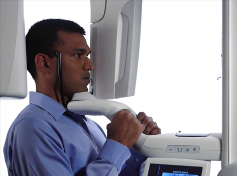
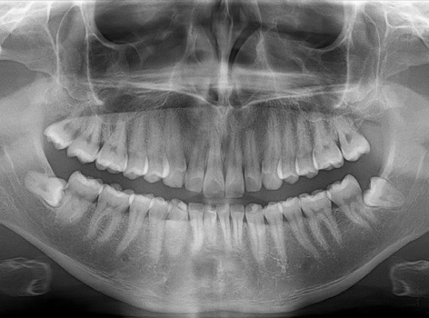
Panoramic x-ray
Panoramic radiography, also called panoramic x-ray, is a two-dimensional (2-D) dental x-ray examination that captures the entire mouth in a single image, including the teeth, upper and lower jaws, surrounding structures and tissues.
A panoramic x-ray is a commonly performed examination by dentists and oral surgeons in everyday practice and is an important diagnostic tool. It covers a wider area than a conventional intraoral x-ray and, as a result, provides valuable information about the maxillary sinuses, tooth positioning and other bone abnormalities. This examination is also used to plan treatment for full and partial dentures, braces, extractions and implants.
A panoramic x-ray can also reveal dental and medical problems such as:
-
Advanced periodontal disease
-
Cysts in the jaw bones
-
Jaw tumors and oral cancer
-
Impacted teeth including wisdom teeth
-
Jaw disorders (also known as temporomandibular joint or TMJ disorders)
-
Sinusitis
In our clinic we use the advanced digital panoramic X-ray equipment ROTOGRAPH EVO which ensures high quality images with the minimum dose of radiation exposed for the patient.
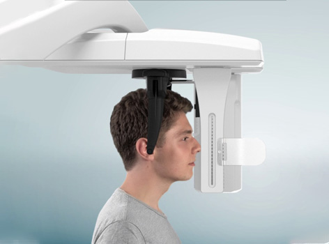
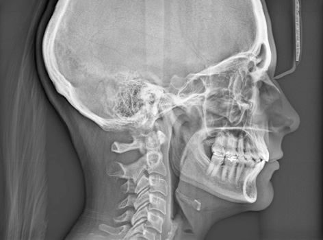
Cephalometric x-ray
A cephalometric x-ray, which is also sometimes referred to simply as a ceph, is a diagnostic radiograph used primarily for orthodontic treatment planning. Once developed, the dentist will use tracing paper, and "trace the ceph" in order to calculate how the patient's jaw and surrounding bone will be affected by orthodontic treatment, along with providing the dentist with a look into the growth pattern of the jaw and teeth. This can be used to determine potential courses of action and routes of treatment.
Cephalometric x-rays are also used by otolaryngologists — doctors who specialize in the treatment of ear, nose, and throat (ENT) disorders such as sleep apnea — because these x-rays provide a view of the patient's airways.
Intraoral x-rays
Intraoral are the most common type of x-ray. There are several types of intraoral x-rays. Each shows different aspects of teeth.
-
Bite-wing x-rays show details of the upper and lower teeth in one area of the mouth. Each bite-wing shows a tooth from its crown (the exposed surface) to the level of the supporting bone. Bite-wing x-rays detect decay between teeth and changes in the thickness of bone caused by gum disease. Bite wing x-rays can also help determine the proper fit of a crown (a cap that completely encircles a tooth) or other restorations (eg, bridges). It can also see any wear or breakdown of dental fillings.
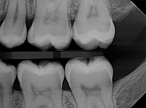
Bitewing without cavities
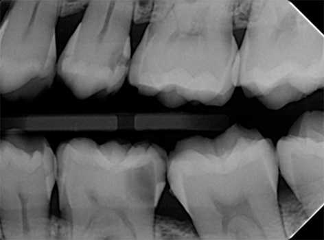
Bitewing with cavities
-
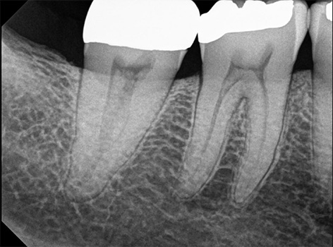
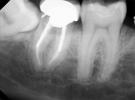
Periapical x-rays show the whole tooth — from the crown, to beyond the root where the tooth attaches into the jaw. Each periapical x-ray shows all teeth in one portion of either the upper or lower jaw. Periapical x-rays detect any unusual changes in the root and surrounding bone structures.
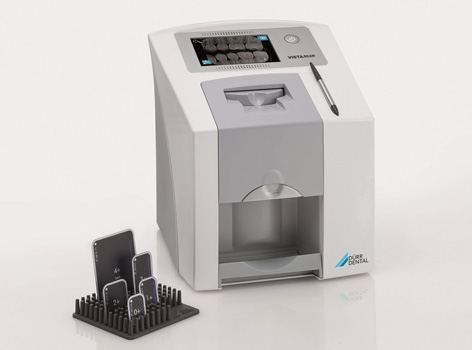
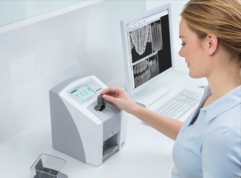
In our dental office we use the VistaScan Mini Easy image plate scanner. It makes image plate diagnostics even faster for dentists. It offers Excellent image quality as well as Rapid image availability.